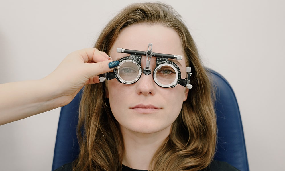

Macular degeneration, often referred to as age-related macular degeneration (AMD), is a progressive eye condition affecting the macula — a tiny central portion of the retina responsible for sharp, central vision. But what does the world look like for those with macular degeneration? This article delves into the symptoms and vision changes experienced by those with AMD.
Understanding the macula
The macula is a pivotal part of the retina, a thin tissue lining the back of the eye. This small, central region, approximately the size of a grain of rice, plays a vital role in our visual perception.
Structure and function
Comprizing millions of light-sensing cells, the macula is densely packed with cone photoreceptors. These are specialized for daylight vision, recognizing colors, and most importantly, detailed central vision. When we focus on something directly in front of us, like the words in a book or intricate details in artwork, it’s the macula that’s hard at work.
Central vs peripheral vision
While the macula is responsible for our sharp central vision, the surrounding part of the retina handles peripheral (side) vision. The crisp and clear images we perceive when looking straight at an object are thanks to the macula, whereas the broader context, or the wider scene around the central focus, is captured by the rest of the retina.
The importance of the macula
Without a healthy macula, tasks requiring precision such as reading, sewing, driving, and recognizing faces become challenging, if not impossible. Such is the importance of this tiny central portion of our eyes.

Types of macular degeneration
Age-related macular degeneration (AMD) mainly occurs in two distinct forms, each with its characteristics and progression patterns.
Dry AMD (Atrophic)
Characteristics: Dry AMD is distinguished by the gradual thinning of the macular tissue. A hallmark sign of this type is the presence of drusen — tiny, yellowish deposits that accumulate under the macula.
Symptoms: Vision loss with dry AMD is generally slow and subtle. Early on, drusen might not cause any symptoms, but as they grow in size and number, they can lead to a dimming or distortion in vision, especially when reading.
Progression: Dry AMD can advance and cause vision loss without turning into wet AMD. An advanced version of dry AMD is called geographic atrophy, where there’s a more noticeable loss of the retinal pigment epithelial cells.

Wet AMD (Neovascular)
Characteristics: Wet AMD is characterized by the growth of abnormal blood vessels from the choroid (a layer behind the retina) under the macula. This is driven by a process called choroidal neovascularization.
Symptoms: These new blood vessels are fragile and tend to leak fluid and blood, leading to scarring of the macula. Consequently, wet AMD can cause rapid and severe vision loss. Early symptoms may include visual distortions where straight lines appear wavy.
Progression: While less common than dry AMD, wet AMD is more aggressive and can lead to significant central vision loss. However, treatments such as anti-VEGF injections can slow the progression and sometimes even improve vision.
It’s worth noting that early-stage dry AMD can progress to late-stage dry AMD or can transform into the wet type. Regular eye check-ups can help in early detection and management of the condition.
Vision with macular degeneration
According to the NHS, “The first symptom is often a blurred or distorted area in your vision.”

“If it gets worse, you might struggle to see anything in the middle of your vision.”

For someone with AMD, central vision becomes increasingly blurred or distorted. The severity and nature of visual symptoms depend on the stage and type of AMD. Here’s what the vision might look like with AMD:
- Blurred vision: One of the earliest signs of AMD is a small blurred area in the center of vision. Over time, this blurred spot might grow larger or become darker.
- Distorted images: Straight lines might appear wavy or bent. For example, door frames and telegraph poles could appear curved.
- Dark, empty areas: As the disease progresses, you might start to see dark or empty areas in the center of your vision. This phenomenon is particularly evident in the advanced stages.
- Color changes: Some individuals report a diminished intensity or brightness of colors.
- Trouble with low light: Those with AMD often find it challenging to adapt to low light levels, making activities in dimly lit environments, like dining in a restaurant, harder.
- Difficulty recognizing faces: Since AMD affects the central vision, recognizing people can become challenging, even if they’re close.
Peripheral vision remains intact
While macular degeneration affects the central vision, one’s peripheral vision remains mostly unaffected. To comprehend this phenomenon, it’s essential to understand the distinction between central and peripheral vision, their functions, and how AMD impacts these areas.
Central vs. peripheral vision
Central vision: This is the vision we use when we focus straight ahead. Governed by the macula, central vision lets us see details clearly and is crucial for tasks like reading, driving, or recognizing faces.
Peripheral vision: On the other hand, peripheral vision encompasses the wide field of view to the sides, above, and below when looking straight ahead. It is not as sharp as central vision but is more sensitive to movement and helps with spatial orientation and mobility.
Why peripheral vision remains unaffected by AMD
Macular degeneration specifically targets the macula, the central portion of the retina. The degradation of the macula leads to the loss of central vision. However, the outer regions of the retina, responsible for peripheral vision, aren’t directly impacted by AMD. This means that even as the central vision becomes increasingly blurred or develops blind spots, the peripheral vision remains largely intact.
Adapting to your new vision
Living with macular degeneration means adjusting to new ways of seeing. People with AMD often rely more on their peripheral vision and might use aids like magnifying glasses and high-contrast reading materials. There’s a focus on maximizing the vision they have left, with many seeking support from low-vision clinics and rehabilitation services.
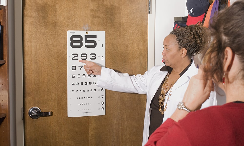
Macular degeneration dramatically alters central vision, making daily activities that require sharp sight more challenging. It’s a journey of adaptation, as individuals learn to navigate the world using their remaining vision. Early detection and intervention can slow the progression of the disease, so regular eye check-ups are essential. If you or someone you know starts to experience any symptoms of AMD, it’s crucial to consult an ophthalmologist promptly.
💡 Further reading: How Long Does It Take To Lose Vision With Macular Degeneration?